Publications
Publications by Theresa Phamduy
Journal articles
Novel digital image analysis using fractal dimension for assessment of skin radiance (2019)
Haeri M, Phamduy T, Cafone N, Turkileri K, Velkov D.
BACKGROUND: Despite a strong desire to quantify skin radiance in the field of cosmetics, there does not exist a robust method to characterize it. Classical shine that quantifies the specular reflection from skin has been commonly used as the metric to characterize radiance. However, it does not always correlate with the perceived radiance as there are many other parameters that inform radiance perception including spatial distribution of shine and color homogeneity.
MATERIALS AND METHODS: In this work, we propose a novel method using fractal analysis to better characterize radiance by considering the spatial heterogeneity of pixel intensities as well as color evenness. A simulated image library (nine images) from very dull to very bright was created using bare face images of 20 panelists. Product images taken post-product usage were ranked along this library by finding the image in the library that most resembles the product image by our algorithm as well as experts. Additionally, classical shine and color measurements were made as benchmarks.
RESULTS: Our results confirm a strong correlation (R2 = 0.99) between the expert radiance rankings and the rankings by fractal dimension algorithm. The new algorithm offers an improved product differentiation compared with classical shine or color measurements.
CONCLUSION: Fractal dimension calculation offers higher sensitivity and resolution compared with other descriptors such as classical shine or color heterogeneity. In cases where the image rank is dominated by pixel intensities rather than color evenness, the image ranks resulting from calculating the fractal dimension is comparable with use of classical shine as the ranking parameter.
Novel 3D and 4D metrics to understand physical tightening mechanisms during eye bag flattening phenomenon (2017)
Phamduy TB, Mercado M, Farran A, Deng Y, Montoya M, Liao I, Langer J, Bouez C, de la Bandera E, Galdi A, Bernard AL, Weinkauf R, Norwood K, Velkov D
Recent advances in skin-tightening products aiming to diminish eye bag puffiness necessitate evaluation methods that quantify complex in vivo surface geometries and soft tissue movement. We answered this need through the development of state-of-the-art methods to measure in vivo eye area geometry in 3D and skin surface motion in 4D (space + time). High-resolution 3D fringe projection scans provided spatial measurements of the facial surface, thus providing information on surface area, zeroed-plane volume, and histogram distribution of vertices distances from the mid-plane. Geometrically, conservation of the surface area and volume indicated that eye bag flattening was driven by surface tightening effects and volume redistribution, rather than true volume loss. After 1-hour of use, the skin-tightening product reshaped the eye area, producing a 19% smoother contour in the eye area closer to the mid-plane. These results correlated with expert visual assessment of the eye bag severity. Furthermore, the mechanism of action was elucidated with parametric analysis of the vertical cross-section profile. Over a 17-minute 4D timelapse capture, the eye bag experienced 12-20% planar contraction strain prior to 8-12% strain at the tear trough. The surface tightening effects stabilized by 15 minutes. Ultimately, these results provide a novel and comprehensive understanding of product mechanism of action, linking dramatic visual improvements to measurable physical phenomena.
Laser Direct-Write onto Live Tissues: A Novel Model for Studying Cancer Cell Migration (2016)
Burks HE, Phamduy TB, Azimi MS, Saksena J, Burow ME, Collins-Burow BM, Chrisey DB, Murfee WL
Investigation into the mechanisms driving cancer cell behavior and the subsequent development of novel targeted therapeutics requires comprehensive experimental models that mimic the complexity of the tumor microenvironment. Recently, our laboratories have combined a novel tissue culture model and laser direct-write, a form of bioprinting, to spatially position single or clustered cancer cells onto ex vivo microvascular networks containing blood vessels, lymphatic vessels, and interstitial cell populations. Herein, we highlight this new model as a tool for quantifying cancer cell motility and effects on angiogenesis and lymphangiogenesis in an intact network that matches the complexity of a real tissue. Application of our proposed methodology offers an innovative ex vivo tissue perspective for evaluating the effects of gene expression and targeted molecular therapies on cancer cell migration and invasion.
Printing cancer cells into intact microvascular networks: a model for investigating cancer cell dynamics during angiogenesis (2015)
Phamduy TB, Sweat RS, Azimi MS, Burow ME, Murfee WL, and Chrisey DB.
While cancer cell invasion and metastasis are dependent on cancer cell–stroma, cancer cell–blood vessel, and cancer cell–lymphatic vessel interactions, our understanding of these interactions remain largely unknown. A need exists for physiologically-relevant models that more closely mimic the complexity of cancer cell dynamics in a real tissue environment. The objective of this study was to combine laser-based cell printing and tissue culture methods to create a novel ex vivo model in which cancer cell dynamics can be tracked during angiogenesis in an intact microvascular network. Laser direct-write (LDW) was utilized to reproducibly deposit breast cancer cells (MDA-MB-231 and MCF-7) and fibroblasts into spatially-defined patterns on cultured rat mesenteric tissues. In addition, heterogeneous patterns containing co-printed MDA-MB-231/fibroblasts or MDA-MB-231/MCF-7 cells were generated for fibroblast-directed and collective cell invasion models. Printed cells remained viable and the cells retained the ability to proliferate in serum-rich media conditions. Over a culture period of five days, time-lapse imaging confirmed fibroblast and MDA-MB-231 cell migration within the microvascular networks. Confocal microscopy indicated that printed MDA-MB-231 cells infiltrated the tissue thickness and were capable of interacting with endothelial cells. Angiogenic network growth in tissue areas containing printed cancer cells was characterized by significantly increased capillary sprouting compared to control tissue areas containing no printed cells. Our results establish an innovative ex vivo experimental platform that enables time-lapse evaluation of cancer cell dynamics during angiogenesis within a real microvascular network scenario.
Spatially-controlled, biomimetic model for breast cancer cell invasion into adipose tissue (in progress, December 2014)
Phamduy TB, Shipman J, Riggs B, Strong AL, Burow ME, Bunnell B, and Chrisey DB
Epithelial-adipose interaction is an integral step in breast cancer cell invasion and progression towards lethal metastatic disease. Understanding the physiological contribution of obesity, a major contributor to breast cancer risk and negative prognosis in post-menopausal patients, on cancer cell invasion requires the detailed co-culture constructs that reflect mammary microarchitecture. We demonstrate the ability to deposit breast cancer cell-ladened hydrogel microbeads into spatially-defined array patterns in hydrogel matrices containing differentiated adipocytes. Laser direct-write, a laser-based bioprinting method, was utilized to target and transfer single microbeads. Z-stack imaging confirmed the three-dimensional nature of the constructs, as well as incorporation of cancer cell-ladened microbeads into the adipose matrix. As an initial validation study, MCF-7 and MDA-MB-231 breast cancer cell invasion was tracked over 2 weeks in the optically-transparent hydrogel scaffold in the presence of differentiated adipocytes obtained from normal weight or obese patient tissue. Thus, our organotypic model successfully integrates fat cells to study cellular and tissue-level interactions towards the early detection of cancer cell invasion into adipose tissue.
'Pioneer' cells at the breast tumor-stroma interface (in progress, DECEMBER 2014)
Phamduy TB, Burks H, Bunnell BA, Burow ME, and Chrisey DB.
Invasion of tumor cells into the surrounding tissue is an integral step in metastasis. Recapitulation of the tumor invasive front requires a mechanistic understanding of spatiotemporal cellular behavior over micrometer-scale protrusion distance and necessitates high-resolution biofabrication tools, such as Laser Direct-Write (LDW), to deposit cells with fine precision. In this review, we debunk the traditional view of the breast tumor-stroma interface, and present the evidence for tumor invasion led by specialized pioneer cancer cells that are driven by intimate interactions with cancer-associated stroma. We address the glaring gap between current co-culture models and physiological relevance due to the absence of sufficient methods to control breast cancer cell placement. To this end, LDW was utilized to generate 2-D, 3-D, and vascularized cancer invasion models with appropriate mammary microarchitecture and stromal context to accurately mimic the tumor microenvironment. An understanding of the spatiotemporal-dependent processes enabling cancer invasion will provide potential diagnostic characteristics and will aid earlier detection of aggressive cancer cell phenotypes.
Laser direct-write of single microbeads into spatially-ordered patterns. (2012)
Phamduy TB, Raof NA, Schiele NR, Yan Z, Corr DT, Huang Y, Xie Y, Chrisey DB.
Biofabrication. 2012 Jun;4(2):025006. doi: 10.1088/1758-5082/4/2/025006
Fabrication of heterogeneous microbead patterns on a bead-by-bead basis promotes new opportunities for sensors, lab-on-a-chip technology and cell-culturing systems within the context of customizable constructs. Laser direct-write (LDW) was utilized to target and deposit solid polystyrene and stem cell-laden alginate hydrogel beads into computer-programmed patterns. We successfully demonstrated single-bead printing resolution and fabricated spatially-ordered patterns of microbeads. The probability of successful microbead transfer from the ribbon surface increased from 0 to 80% with decreasing diameter of 600 to 45 µm, respectively. Direct-written microbeads retained spatial pattern registry, even after 10 min of ultrasonication treatment. SEM imaging confirmed immobilization of microbeads. Viability of cells encapsulated in transferred hydrogel microbeads achieved 37 ± 11% immediately after the transfer process, whereas randomly-patterned pipetted control beads achieved a viability of 51 ± 25%. Individual placement of >10 µm diameter microbeads onto planar surfaces has previously been unattainable. We have demonstrated LDW as a valuable tool for the patterning of single, micrometer-diameter beads into spatially-ordered patterns.
Suppression of triple-negative breast cancer metastasis by pan-DAC inhibitor panobinostat via inhibition of ZEB family of EMT master regulators (2014)
Rhodes LV, Tate CR, Segar HC, Burks HE, Phamduy TB, Hoang V, Elliott S, Gilliam D, Pounder FN, Anbalagan M, Chrisey DB, Rowan BG, Burow ME, Collins-Burow BM.
Breast Cancer Res Treat. 2014 Jun;145(3):593-604. doi: 10.1007/s10549-014-2979-6.
Triple-negative breast cancer (TNBC) is a highly aggressive breast cancer subtype that lacks effective targeted therapies. The epithelial-to-mesenchymal transition (EMT) is a key contributor in the metastatic process. We previously showed the pan-deacetylase inhibitor LBH589 induces CDH1 expression in TNBC cells, suggesting regulation of EMT. The purpose of this study was to examine the effects of LBH589 on the metastatic qualities of TNBC cells and the role of EMT in this process. A panel of breast cancer cell lines (MCF-7, MDA-MB-231, and BT-549), drugged with LBH589, was examined for changes in cell morphology, migration, and invasion in vitro. The effect on in vivo metastasis was examined using immunofluorescent staining of lung sections. EMT gene expression profiling was used to determine LBH589-induced changes in TNBC cells. ZEB overexpression studies were conducted to validate requirement of ZEB in LBH589-mediated proliferation and tumorigenesis. Our results indicate a reversal of EMT by LBH589 as demonstrated by altered morphology and altered gene expression in TNBC. LBH589 was shown to be a more potent inhibitor of EMT than other HDAC inhibitors, SAHA and TMP269. Additionally, we found that LBH589 inhibits metastasis of MDA-MB-231 cells in vivo. These effects of LBH589 were mediated in part by inhibition of ZEB, as overexpression of ZEB1 or ZEB2 mitigated the effects of LBH589 on MDA-MB-231 EMT-associated gene expression, migration, invasion, CDH1 expression, and tumorigenesis. These data indicate therapeutic potential of LBH589 in targeting EMT and metastasis of TNBC.
Laser Direct-Write of Embryonic Stem Cells and Cells Encapsulated in Alginate Beads for Engineered Biological Constructs (2012)
Phamduy TB, Dias AD, Abdul Raof N, Schiele NR, Corr DT, Xie Y, and Chrisey DB
MRS Proceedings / Volume 1418 / 2012 / DOI: http://dx.doi.org/10.1557/opl.2012.798
The ability to control the deposition of mouse embryonic stem cells (mESCs), and mESCs encapsulated in 200-μm diameter alginate microbeads, into customized patterns has recently been achieved using laser direct-write (LDW). Gelatin-based LDW was utilized to target and reproducibly deposit groups of cells directly onto receiving substrate surfaces. Live/dead staining for cell viability and immunocytochemistry for the pluripotency marker, Oct-4, indicated that transferred mESCs were viable following transfer, and maintained an important embryonic stem cell marker, respectively. LDW was further used to print mESCs encapsulated in hydrogel microbeads into customized patterns on a single-bead basis. Recent efforts have also achieved patterns of discrete co-cultures of mESCs and breast cancer cells in separate hydrogel microbeads. Altogether, we demonstrated the feasibility of LDW to print patterns of mESCs and mESC-microbeads for the biomimetic assembly of engineered cellular constructs and tissue models.
Impedance spectroscopy for the detection of unknown toxins
Riggs B, Plopper G, Phamduy TB, Paluh J, Corr DT, and Chrisey DB
Proceedings of SPIE. 8371A, 49
Advancements in biological and chemical warfare has allowed for the creation of novel toxins necessitating a universal, real-time sensor. We have used a function-based biosensor employing impedance spectroscopy using a low current density AC signal over a range of frequencies (62.5 Hz-64 kHz) to measure the electrical impedance of a confluent epithelial cell monolayer at 120 sec intervals. Madin Darby canine kidney (MDCK) epithelial cells were grown to confluence on thin film interdigitated gold electrodes. A stable impedance measurement of 2200 Ω was found after 24 hrs of growth. After exposure to cytotoxins anthrax lethal toxin and etoposide, the impedance decreased in a linear fashion resulting in a 50% drop in impedance over 50hrs showing significant difference from the control sample (~20% decrease). Immunofluorescent imaging showed that apoptosis was induced through the addition of toxins. Similarities of the impedance signal shows that the mechanism of cellular death was the same between ALT and etoposide. A revised equivalent circuit model was employed in order to quantify morphological changes in the cell monolayer such as tight junction integrity and cell surface area coverage. This model showed a faster response to cytotoxin (2 hrs) compared to raw measurements (20 hrs). We demonstrate that herein that impedance spectroscopy of epithelial monolayers serves as a real-time non-destructive sensor for unknown pathogens.
Evaluation of electric cell-substrate impedance sensing for the detection of nanomaterial toxicity
McAuley E, Mohanraj B, Phamduy TB, Plopper GE, Corr DT, and Chrisey DB
Int. J. of Biomedical Nanoscience and Nanotechnology, 2011 Vol.2, No.2, pp.136 - 151
Electric cell-substrate impedance sensing (ECIS) is an in situ and real-time monitoring system used to detect toxic agents by monitoring changes in impedance of a confluent cell monolayer. When toxic agents are introduced to cells, they can cause a change in the cell barrier function, a direct measure of the resistance to current flow caused by tight junction formation between cells. This exposure results in an immediate, quantitative change in the measured resistance between the electrodes, thus, continuously monitoring cell behaviour and by extension, toxic exposure. We have developed an ECIS-based protocol to functionally characterise epithelial cell response when challenged by different toxicants, particularly silver and copper nanoparticles. We verified our impedance changes with observed structural changes by fluorescent staining of zonula occludens-1 (ZO-1) protein in the tight junctions of a model epithelial cell line.
PD-L1 interacts specifically with B7-1 to regulate T cell function.
Butte MJ, Keir ME, Phamduy TB, Sharpe AH, and Freeman GJ.
The Journal of Immunology, 2007. 178, 88.24
Pathways in the B7:CD28 family of costimulatory molecules regulate T cell activation and tolerance. B7-dependent responses in Cd28(-/-)Ctla4(-/-) T cells together with reports of stimulatory and inhibitory functions for Programmed Death-1 Ligand 1 or 2 molecules (PD-L1 or PD-L2) have suggested additional receptors for these B7 family members. We show that B7-1 and PD-L1 interacted with affinity intermediate to that of B7-1:CD28 and B7-1:CTLA-4. The PD-L1:B7-1 interface overlapped with the B7-1:CTLA-4 and PD-L1:PD-1 (Programmed Death-1) interfaces. T cell activation and cytokine production were inhibited by the interaction of B7-1 with PD-L1. The responses of PD-1-deficient versus PD-1,B7-1 double-deficient T cells to PD-L1 and of CD28,CTLA-4 double-deficient versus CD28,CTLA-4,PD-L1 triple-deficient T cells to B7-1 demonstrated that PD-L1 and B7-1 interact specifically to inhibit T cell activation. Our findings point to a substantial bidirectional inhibitory interaction between B7-1 and PD-L1 and add an additional dimension to immunoregulatory functions of the B7:CD28 family.
Book chapters
The Power of CAD/CAM Laser Bioprinting at the Single Cell Level: Evolution of Printing (in progress, 2014)
Sklare S, Phamduy TB, Curley JL, Huang Y, and Chrisey DB.
3D Bioprinting and Nanotechnology in Tissue Engineering. Elsevier.
Singlecell deposition onto non-restrictive two-dimensional (2D) and into three-dimensional (3D) environments with high accuracy and reproducibility is not only possible, but entering a new stage of maturity by way of systems engineering and a critical mass of past iterations. Coupling computer-aided design (CAD) and manufacturing (CAM) principlesto laser systems enables researchers to create reproducible biological models with single-cell precision. In this chapter, we illustrate the importance of single-cell deposition and introduce laser-assisted bioprinting as a viable method of printing individual cells. In particular, Matrix-Assisted Pulsed-Laser Evaporation Direct Write (MAPLE-DW), a bottom-up biofabrication technique, will be used to showcase the advantages of laser-assisted, nozzle-free, and contactless printing methods, while avoiding stochastic printing methods. Currently in its third iteration, recent improvements to MAPLE-DW focused on potential scalability, single cell deposition and ease of use.
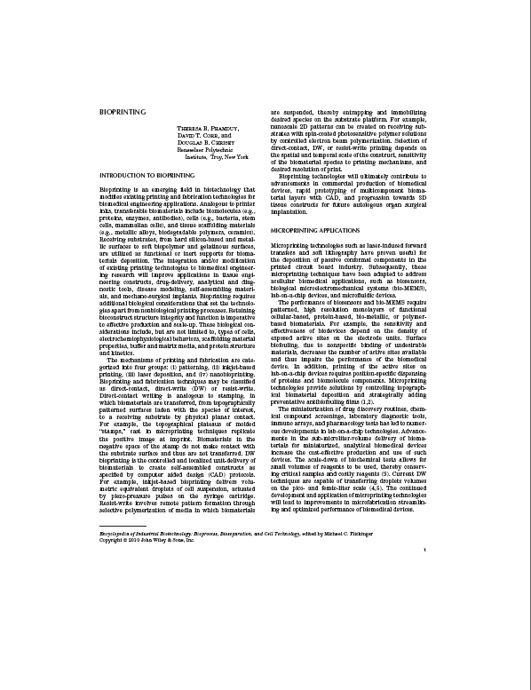
Bioprinting
Phamduy TB, Corr DT, and Chrisey DB.
Encyclopedia of Industrial Biotechnology. Wiley. 2010.
Bioprinting refers to the spatially-controlled deposition of disparate biomaterials that is carried out by various printing techniques for the bottom-up transfer of cells, biomolecules, and scaffold. The applications and techniques for bioprinting discussed in this chapter address the emergent demands of sub-micrometer patterns for fundamental study, device construction, and bottom-up tissue engineering. The advantages of bioprinting stem from its unique attributes of controllability, automated deposition, CAD/CAM construction, bottom-up writing, and rapid prototyping. As advancements in biotechnology gravitate towards the micro-scale and nano-scale, precise deposition of biomaterial patterns becomes critical for many applications, including biomedical devices. In addition, the drive for the rapid prototyping of tissue constructs requires the combination of computer aided design and high-resolution repeatability that bioprinting is able to achieve. Examples of bioprinting methods include patterning, ink-jet based bioprinting, laser deposition, and nanobioprinting. Bioprinting is poised to be a significant tool for metric transfers of fragile biomaterials in both biomedical engineering research and industrial biotechnology applications.


